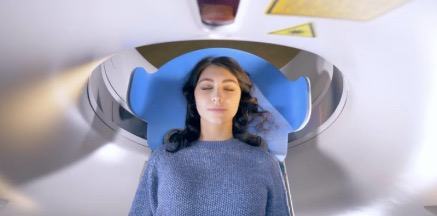
Premier Diagnostic Imaging is a freestanding medical imaging facility with medical and diagnostic exam services offering state-of-the-art medical screening and diagnostic examinations at a low cost. After your examination, one of our radiologists will analyze your images and send a signed report with his interpretation to your primary care physician. Your physician’s office will inform you of your results, and you may also log in to our patient portal to access the report and images from your examination. Select the specific medical and diagnostic exam services you need below to learn more about how to prepare and what to expect during an examination.

CT, sometimes called CAT scanning, uses special x-ray equipment to obtain images from different angles, which are then processed by computer to show a cross-section of body tissues and organs. There are many applications for CT scanning, including assessing injury and diagnosing disease. Some screening exams that use the CT scanner are as follows:
Women should begin receiving mammograms every year after age 40, according to the American College of Radiology. Statistics indicate that one in eight women will develop breast cancer sometime in her lifetime. Premier’s 3D mammograms provide exceptionally sharp images designed to detect breast cancer early, and our SmartCurve plates provide a more comfortable experience.
Ultrasound imaging, or sonography, is a method of seeing inside the human body through the use of high-frequency sound waves. Because ultrasound images are captured in real-time, they can show movement of internal tissues and organs.
MRI uses radio waves, a strong magnetic field and a computer to generate detailed, cross-sectional images of human anatomy. Because it produces better tissue images than x-rays, MRI is most commonly used for the brain, spine, thorax, vascular system, and musculoskeletal system (including the knee and ankle).
Digital x-ray (radiography) uses a very small dose of ionizing radiation to produce pictures of the body’s internal structures. X-rays are the oldest and most frequently used form of medical imaging. Fluoroscopy studies moving body structures, so you can think if it as an x-ray movie.
Bone density testing (DEXA) is the only way to detect low bone mass. Osteoporosis is a disease in which bones become fragile and more likely to break. It is recommended that all women over 65 receive a DEXA bone density scan.
Pain management injections provide temporary relief from back pain, lasting from days to months, reducing the inflammation and swelling. This can allow your condition to improve with physical therapy and exercise.
Biopsies take a small sample of cells from a suspicious area within the body and examine them under a microscope to determine a diagnosis.